Two-class microscopy segmentation with 2D U-Net
In this tutorial we will learn the following features:
Training of a segmentation model (U-Net 2D) with two-class labels on a single contrast on microscopy PNG images,
Testing of a trained model and computation of evaluation metrics,
Visualization of the outputs of a trained model.
Download dataset
We will use a publicly available dataset consisting of 10 microscopy samples of rat spinal cord.
To download the dataset (~11MB), run the following command in your terminal:
# Download data
ivadomed_download_data -d data_axondeepseg_sem
Configuration file
In ivadomed, training is orchestrated by a configuration file. Examples of configuration files are available in
the ivadomed/config/ and the documentation is available in Configuration File.
In this tutorial, we will use the configuration file: ivadomed/config/config_microscopy.json.
First off, copy this configuration file in your local directory (to avoid modifying the source file):
cp <PATH_TO_IVADOMED>/ivadomed/config/config_microscopy.json .
Then, open it with a text editor. Below we will discuss some of the key parameters to perform a two-class 2D microscopy segmentation training.
command: Action to perform. Here, we want to train a model, so we set the fields as follows:"command": "train"path_output: Folder name that will contain the output files (e.g., trained model, predictions, results)."path_output": "log_microscopy_sem"loader_parameters:path_data: Location of the dataset. As discussed in Data, the dataset should conform to the BIDS standard. Modify the path so it points to the location of the downloaded dataset."path_data": ["data_axondeepseg_sem"]loader_parameters:target_suffix: Suffix of the ground truth segmentations. The ground truths are located under thedata_axondeepseg_sem/derivatives/labelsfolder. In our case, the suffix are_seg-axon-manualand_seg-myelin-manual:"target_suffix": ["_seg-axon-manual", "_seg-myelin-manual"]
loader_parameters:extensions: List of file extensions of the microscopy data. In our case, both the raw data and derivatives are “.png” files."extensions": [".png"]loader_parameters:contrast_params: Contrast(s) of interest. In our case, we are training a single contrast model with contrastSEM."contrast_params": { "training_validation": ["SEM"], "testing": ["SEM"], "balance": {} }
loader_parameters:slice_axis: Orientation of the 2D slice to use with the model. 2D PNG files must use defaultaxial."slice_axis": "axial"split_dataset:split_method: Describe the metadata used to split the train/validation/test sets. Here,sample_idfrom thesamples.tsvfile will shuffle all samples, then split them between train/validation/test sets.split_dataset:train_fraction: Fraction of the dataset’ssample_idin the train set. In our case0.6.split_dataset:test_fraction: Fraction of the dataset’ssample_idin the test set. In our case0.1."split_method" : "sample_id" "train_fraction": 0.6 "test_fraction": 0.1
training_parameters:training_time:num_epochs: The maximum number of epochs that will be run during training. Each epoch is composed of a training part and a validation part. It should be a strictly positive integer. In our case, we will use 50 epochs."num_epochs": 50default_model:length_2D: Size of the 2D patches used as model’s input tensors. We recommend using patches between 256x256 and 512x512. In our case, we use patches of 256x256.default_model:stride_2D: Pixels’ shift over the input matrix to create 2D patches. In our case, we use a stride of 244 pixels in both dimensions, resulting in an overlap of 12 pixels between patches."length_2D": [256, 256] "stride_2D": [244, 244]
postprocessing:binarize_maxpooling: Used to binarize predictions across all classes in multiclass models. For each pixel, the class, including the background class, with the highest output probability will be segmented."binarize_maxpooling": {}evaluation_parameters:object_detection_metrics: Used to indicate if object detection metrics (lesions true positive rate, lesions false detection rate and Hausdorff score) are computed or not at evaluation time. For the axons and myelin segmentation task, we set this parameter tofalse."object_detection_metrics": falsetransformation:Resample: Used to resample images to a common resolution (in mm) before splitting into patches, according to each image real pixel size. In our case, we resample the images to a common resolution of 0.0001 mm (0.1 μm) in both dimensions."Resample": { "hspace": 0.0001, "wspace": 0.0001 },
Train model
Once the configuration file is ready, run the training:
ivadomed -c config_microscopy.json
Alternatively, the “command”, “path_output”, and “path_data” arguments can be passed as CLI flags in which case they supersede the configration file, see Usage.
ivadomed --train -c config_microscopy.json --path-data path/to/bids/data --path-output path/to/output/directory
Note
If a compatible GPU is available, it will be used by default. Otherwise, training will use the CPU, which will take a prohibitively long computational time (several hours).
The main parameters of the training scheme and model will be displayed on the terminal, followed by the loss value on training and validation sets at every epoch. To know more about the meaning of each parameter, go to Configuration File. The value of the loss should decrease during the training.
No CLI argument given for command: ( --train | --test | --segment ). Will check config file for command...
CLI flag --path-output not used to specify output directory. Will check config file for directory...
CLI flag --path-data not used to specify BIDS data directory. Will check config file for directory...

Creating output path: log_microscopy_sem
Using GPU ID 0

Selected architecture: Unet, with the following parameters:
dropout_rate: 0.2
bn_momentum: 0.1
depth: 4
is_2d: True
final_activation: sigmoid
length_2D: [256, 256]
stride_2D: [244, 244]
folder_name: model_seg_rat_axon-myelin_sem
in_channel: 1
out_channel: 3

Dataframe has been saved in log_microscopy_sem/bids_dataframe.csv.
After splitting: train, validation and test fractions are respectively 0.6, 0.3 and 0.1 of sample_id.

Selected transformations for the ['training'] dataset:
Resample: {'hspace': 0.0001, 'wspace': 0.0001}
RandomAffine: {'degrees': 2.5, 'scale': [0.05, 0.05], 'translate': [0.015, 0.015], 'applied_to': ['im', 'gt']}
ElasticTransform: {'alpha_range': [100.0, 150.0], 'sigma_range': [4.0, 5.0], 'p': 0.5, 'applied_to': ['im', 'gt']}
NormalizeInstance: {'applied_to': ['im']}
Selected transformations for the ['validation'] dataset:
Resample: {'hspace': 0.0001, 'wspace': 0.0001}
NormalizeInstance: {'applied_to': ['im']}

Loading dataset: 100%|████████████████████████████████████████████████████████████████| 3/3 [00:00<00:00, 738.48it/s]
Loaded 76 axial patches of shape [256, 256] for the validation set.
Loading dataset: 100%|████████████████████████████████████████████████████████████████| 6/6 [00:00<00:00, 829.21it/s]
Loaded 252 axial patches of shape [256, 256] for the training set.
Creating model directory: log_microscopy_sem/model_seg_rat_axon-myelin_sem

Initialising model's weights from scratch.
Scheduler parameters: {'name': 'CosineAnnealingLR', 'base_lr': 1e-05, 'max_lr': 0.01}

Selected Loss: DiceLoss
with the parameters: []
Epoch 1 training loss: -0.6894.
Epoch 1 validation loss: -0.7908.
After 50 epochs (see "num_epochs" in the configuration file), the Dice score on the validation set should be ~85%.
Note
When loading the images for training or evaluation, a temporary NIfTI file will be created for each images in the
dataset directory (path_data) alongside the original PNG files.
Evaluate model
To test the trained model on the testing sub-dataset and compute evaluation metrics, run:
ivadomed -c config_microscopy.json --test
If you prefer to use config files over CLI flags, set “command” to the following in you config file:
"command": "test"
Then run:
ivadomed -c config_microscopy.json
The model’s parameters will be displayed in the terminal, followed by a preview of the results for each image.
The resulting segmentations are saved for each image in the <PATH_TO_OUT_DIR>/pred_masks while a CSV file,
saved in <PATH_TO_OUT_DIR>/results_eval/evaluation_3Dmetrics.csv, contains all the evaluation metrics.
For more details on the evaluation metrics, see ivadomed.metrics.
CLI flag --path-output not used to specify output directory. Will check config file for directory...
CLI flag --path-data not used to specify BIDS data directory. Will check config file for directory...

Output path already exists: log_microscopy_sem
Using GPU ID 0

Selected architecture: Unet, with the following parameters:
dropout_rate: 0.2
bn_momentum: 0.1
depth: 4
is_2d: True
final_activation: sigmoid
length_2D: [256, 256]
stride_2D: [244, 244]
folder_name: model_seg_rat_axon-myelin_sem
in_channel: 1
out_channel: 3

Dataframe has been saved in log_microscopy_sem/bids_dataframe.csv.
After splitting: train, validation and test fractions are respectively 0.6, 0.3 and 0.1 of sample_id.

Selected transformations for the ['testing'] dataset:
Resample: {'hspace': 0.0001, 'wspace': 0.0001}
NormalizeInstance: {'applied_to': ['im']}

Loading dataset: 100%|████████████████████████████████████████████████████████████████| 1/1 [00:00<00:00, 413.48it/s]
Loaded 16 axial patches of shape [256, 256] for the testing set.
Loading model: log_microscopy_sem/best_model.pt

Inference - Iteration 0: 100%|████████████████████████████████████████████████████████████████| 4/4 [00:01<00:00,  2.89it/s]
Lossy conversion from float64 to uint8. Range [0, 1]. Convert image to uint8 prior to saving to suppress this warning.
Lossy conversion from float64 to uint8. Range [0, 1]. Convert image to uint8 prior to saving to suppress this warning.
{'dice_score': 0.8381376827003003, 'multi_class_dice_score': 0.8422281034034607, 'precision_score': 0.8342335786851753,
'recall_score': 0.8420784999205466, 'specificity_score': 0.9456594910680598, 'intersection_over_union': 0.7213743575471384,
'accuracy_score': 0.9202670087814067, 'hausdorff_score': 0.0}

Run Evaluation on log_microscopy_sem/pred_masks

Evaluation: 100%|████████████████████████████████████████████████████████████████| 1/1 [00:13<00:00, 13.56s/it]
Lossy conversion from float64 to uint8. Range [0.0, 3.0]. Convert image to uint8 prior to saving to suppress this warning.
Lossy conversion from float64 to uint8. Range [0.0, 3.0]. Convert image to uint8 prior to saving to suppress this warning.
                             avd_class0  avd_class1  dice_class0  dice_class1  ...  vol_gt_class0  vol_gt_class1  vol_pred_class0  vol_pred_class1
image_id
sub-rat3_sample-data9_SEM    0.082771    0.082971    0.868964     0.815492     ...  1.256960e-07   1.574890e-07   1.152920e-07     1.705560e-07

[1 rows x 26 columns]
The test image segmentations are stored in <PATH_TO_OUT_DIR>/pred_masks/ in PNG format and have the same name as
the input image with the suffix <class-index>_pred.png. In our case: sub-rat3_sample-data9_SEM_class-0_pred.png and
sub-rat3_sample-data9_SEM_class-1_pred.png for axons and myelin respectively (in the same order as target_suffix).
A temporary NIfTI files containing the predictions for both classes with the suffix _pred.nii.gz will also be
present.
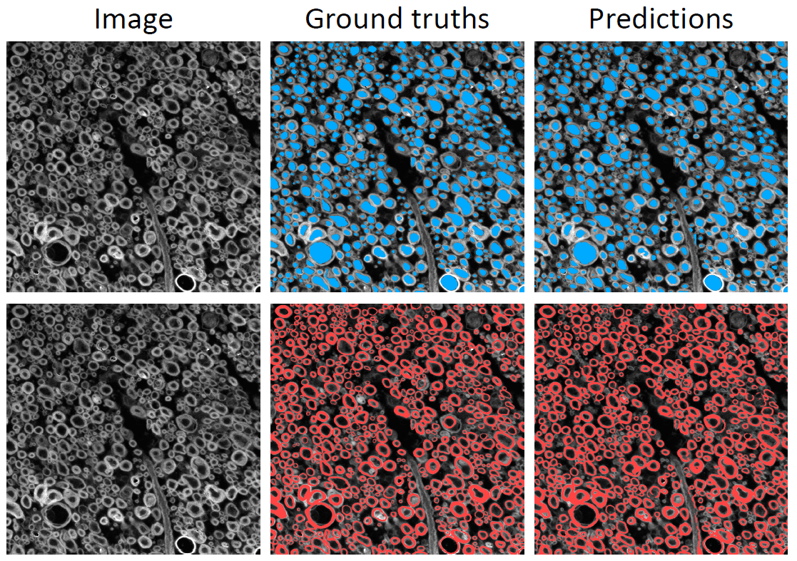
After the training for 50 epochs, the segmentations should be similar to the one presented in the following image.
The ground truth segmentations and predictions of the axons and myelin are presented in blue and red respectively for
sub-rat3_sample-data9_SEM):
Another set of test image segmentations are also present in <PATH_TO_OUT_DIR>/pred_masks/ with the suffix _pred-TP-FP-FN when the evaluation_parameters:object_detection_metrics is set to true. These files include 3 possible values depending if each object detected in the prediction compared to the ground-truth is a True Positive (TP), False Positive (FP) or False Negative (FN). In PNG files (.png), the respective values for TP, FP and FN are 85, 170 and 255.